PULLMAN, WA – With the addition of a new CT scanner, Washington State University’s Veterinary Teaching Hospital can deliver faster, more precise diagnoses — improving the outcomes for animals across the Inland Northwest.
The hospital recently upgraded to Canon’s high-performance Aquilion Prime S80 system from the previous scanner installed in 2010. The new scanner, which is now in operation after several weeks in which the hospital operated without the diagnostic imaging service, features 80 image slices per rotation. This higher spatial resolution provides clearer, more detailed images than the previous 16-slice system, which was well suited for routine imaging but more limited for advanced diagnostics.
“This new CT represents a major advancement in imaging capability, efficiency, and patient safety, allowing our veterinarians to diagnose complex conditions with greater accuracy while reducing radiation exposure for our animal patients, and it was made possible entirely by our hospital’s private donors,” hospital Director Dr. Raelynn Farnsworth said.
CT, or computed topography, works by capturing X-ray images taken at high speed as they rotate around a subject. These individual X-rays are used to recreate detailed 3D images of organs, bones, and tissues that can help doctors in human and veterinary medicine provide the most accurate medical advice.
One of the most significant improvements is the new scanner’s ability to take high-quality images at low-dose radiation, which, in comparison to the previous machine, will reduce radiation exposure for patients.
In addition, faster rotation and reconstruction speeds — up to 60 images per second — will improve workflow and make the system especially valuable for emergency cases and other time-sensitive or complex studies.
“That means we will be able to help more patients in a shorter amount of time,” Farnsworth said.
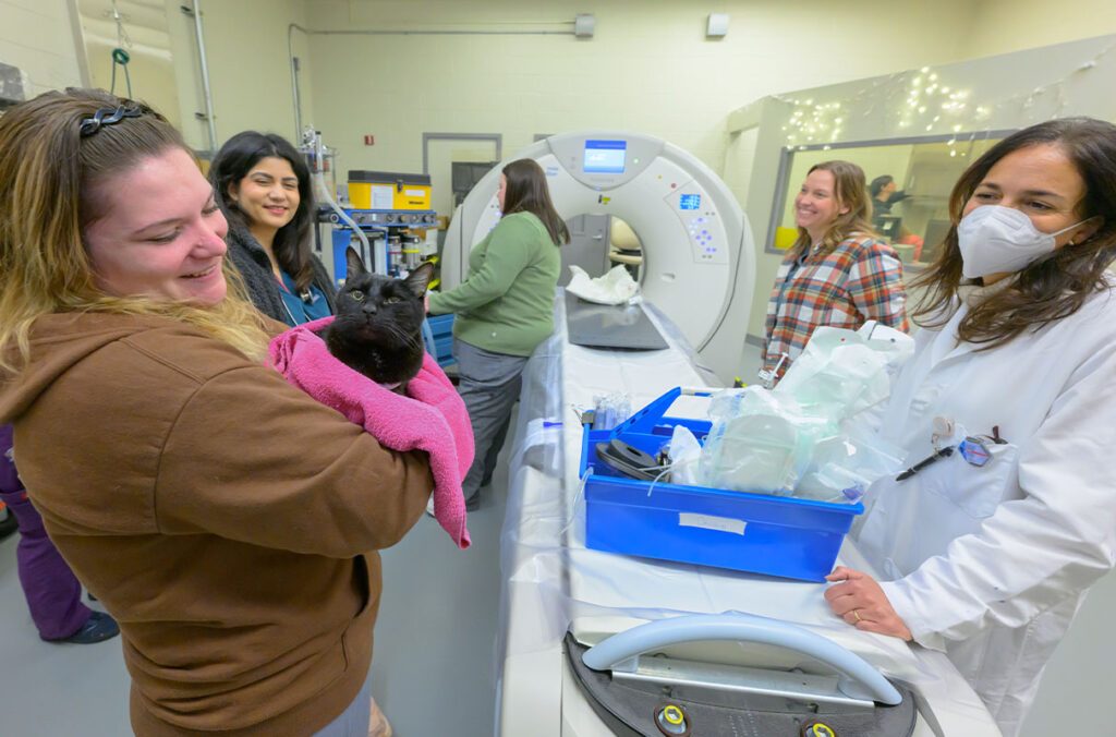
The new CT is already providing critical medical information for animals like Tux, a 14-year-old tuxedo cat that was the first patient to be scanned with the new CT.
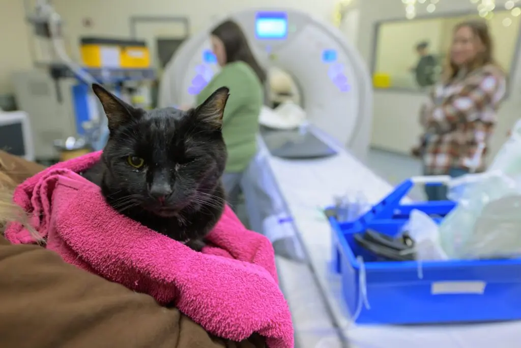
A beloved member of the Crouch family in Spokane, Tux came to WSU the way many animals do — after being referred by their primary veterinarian.
“He had this cough that reminded me of when they have a hairball, but we weren’t finding any hairballs, so we took him to his normal vet,” Meggie Crouch, Tux’s owner, said.
A single X-ray of Tux’s chest revealed a lump in one of his lungs, which prompted a visit to WSU for an ultrasound and later a CT scan.
“The CT was really helpful diagnostically because it helped us know there weren’t any other masses that were this large, but it also showed there were other small unknown changes to his lungs,” Crouch said. “I know it doesn’t seem like it, but that was good information for us because a follow-up CT will show exactly what has changed.”
Tux will return in late February for his follow-up scan, but Crouch said he is already showing signs he’s on the mend, and she couldn’t be more grateful for WSU.
“It was really nice to have the option of going down to WSU for a CT because our other option was to go to a place that mostly does humans, so I felt like going to WSU was going to be the best for us, not only because of the new technology but also because of the care and compassion that everyone puts into our pets,” Crouch said.